Comprehensive List of Researchers "Information Knowledge"
Department of Media Science
- Name
- MORI, Kensaku
- Group
- Joint Member
- Title
- Professor
- Degree
- Dr. of Engineering
- Research Field
- Image processing / Visualization / Medical image processing

Current Research
Development of Image Processing Techniques and Applications to Medical Images
OUTLINESuch medical imaging devices as CT or MR scanners, which are used in hospitals for visualizing the inside of human bodies, have made remarkable progress. Today it is possible to take very precise volumetric images of a wide area of a human body in twenty seconds. We are investigating systems that enable the following: (a) the navigation of the inside of a human body (diagnosis of images), (b) finding positions where abnormal regions can be seen, (c) image deformation if needed, and (d) assisting or guiding surgery and examinations (guidance-based on images). We are focusing on a virtual endoscopy system that enables us to navigate the inside of a human based on 3D or 4D medical images. This virtual endoscopy system is utilized for computer-aided diagnosis for medical images and computer-aided surgery. We are also developing fundamental techniques, including image recognition and understanding, computer vision, computer graphics, and user interfaces.
TOPICS
(1) Virtual endoscopy system
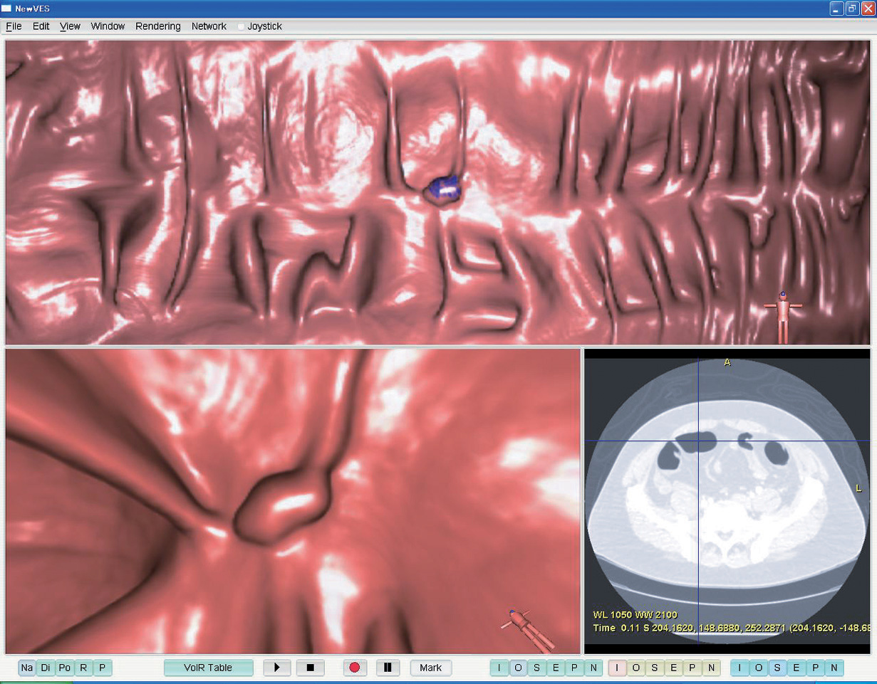
The virtual endoscopy system is a system that can generate endoscopic-like images based on 3D or higher medical images. We have developed techniques for the segmentation of organ regions from 3D medical images and interactive visualization. A virtual endoscopy system has several advantages: (a) no pain to patients, (b) observation from arbitrary viewpoints and directions, and (c) observation of anatomical structures existing beyond the organ wall currently observed by employing semi-translucent display, compared to a real endoscope such as a bronchoscope or colonoscope. The virtual endoscopy system developed here is now widely being used as a new observation tool for medical images in the clinical field. We are now extending the it to be a colonic polyp inspection system and are developing fundamental techniques related to it.
(2) Endoscopy navigation system
In this research project, we are developing endoscopy navigation systems that guide physicians during examinations or surgery using an endoscope. We use medical images taken before examinations as a kind of “road map.” We display the current location of an endoscope, the locations of important organs, and paths to target points by analyzing endoscopic images. This system resembles a GPS navigation system for vehicles. By utilizing this system in a real clinical field, a physician can make appropriate decisions during endoscopy or surgery. In the case of bronchoscopy, it is impossible to sense bronchoscope position and orientation by sensors due to special limitations. We are developing a method to estimate bronchoscope motion based on image registration between real bronchoscopic images and virtual bronchoscopic images derived from CT images.
(3) Image deformation, computer-aided diagnosis, and computer-aided surgery
We are developing a method to virtually stretch or deform organs for medical image diagnosis or surgical planning by extracting target organ regions from 3D medical images and modeling them as elastic objects. By deforming and visualizing them, we can generate virtually stretched views of the stomach or the abdomen. Images generated by this method are quite useful for diagnosis and surgical planning.
FUTURE WORK
We are planning to make these methods widely usable in real clinical fields for developing next-generation medical engineering. Fundamental techniques will also be developed for constructing advanced medical image processing devices.

Figure : CAD system for colonic polyp detection
Career
- Kensaku Mori received a Dr. of Engineering degree in Information Engineering from Nagoya University in 1996.
- He was a research and a postdoc fellow of JSPS from 1994-1996 and a visiting assoc. prof. at Stanford University from 2001 to 2002.
- Since 2003, he has been an assoc. prof. of the Graduate School of Information Science, Nagoya University.
Academic Societies
- IEEE
- ACM
- ISCAS
- IEICE
- IPSJ
- JSBME
- JAMIT
- CADM
- JSCAS
Publications
- A method for bronchoscope tracking by combining a position sensor and image registration, Computer Aided Surgery, 37(1), pp. 109-117 (2006).
- Fast Generation of Digitally Reconstructed Radiographs Using Attenuation Fields With Application to 2D-3D Image Registration, IEEE Trans. MI, 24(11), pp. 1441-1454 (2005).
- Tracking of a bronchoscope using epipolar geometry analysis and intensity-based image registration of real and virtual endoscopic images, Med. Img. Ana., 6, pp. 321-336 (2002).


